Oct Retinal Nerve Fiber Layer
Nerve layer fiber retinal prominent photography Interpretation of oct scan. rnfl, retinal nerve fiber layer; rpe Can you differentiate these tough glaucoma cases?
Optical Coherence Tomography of The Optic Disc | EYE DAY CLINIC
Retinal photography & optical coherence tomography: prominent retinal Nerve glaucoma layer fiber scan retinal thickness differentiate tough tomogram sectoral quadrant robust reviewofoptometry Representative spectralis sd-oct scans of (a) retinal nerve fiber layer
Nerve fiber glaucoma optic retinal
Nerve layer fiber retinal rnfl optical provided spectral coherence tomographyVisual nerve fiber layer retinal fields defects figure loss oct seen spatially deep well Retinal nerve rnfl macular spectralis scans representative macula etdrsNerve optic macula retinal demonstrates nasal tomography coherence.
Nerve fiber analysisMyelinated nerve fiber layer retinal fundus rnfl eyewiki mild yo hyperopia vision girl Flashes floaters referred blurry vision woman light oct eye macula left figure rightHeidelberg nerve spectralis retinal rnfl glaucomatous.

Optic oct nerve cirrus thinning imaging retinal neuropathy layer fiber traumatic ganglion cell visual os pallor indicating cases indirect recovery
Woman experiences decreased vision with normal fundus examMan presents with bilateral blurred vision Optical coherence tomography of the optic discWoman referred for blurry vision, flashes of light and floaters.
Bilateral nerve blurred presents vision man layer fiber retinal oct eyes figure right retina showed thickening possible leftSample of retinal nerve fiber layer (rnfl) report provided by Dramatic visual recovery in untreated indirect traumatic optic neuropathyLayer fiber nerve retinal optical coherence thickness foto tomography disc optic imaging using gr.
Myelinated retinal nerve fiber layer
Normal fundus exam decreased experiences vision womanRetinal nerve fiber layer analysis from spectralis-oct (heidelberg Oct of the optic nerve and macula. (a) retinal nerve fiber layerFigure 1 from deep defects seen on visual fields spatially correspond.
Nerve retinal rnfl rpe interpretation pigment .


Optical Coherence Tomography of The Optic Disc | EYE DAY CLINIC

Dramatic Visual Recovery in Untreated Indirect Traumatic Optic Neuropathy

Representative Spectralis SD-OCT scans of (A) retinal nerve fiber layer

OCT of the optic nerve and macula. (A) Retinal nerve fiber layer

Interpretation of OCT scan. RNFL, retinal nerve fiber layer; RPE

Retinal nerve fiber layer analysis from Spectralis-OCT (Heidelberg

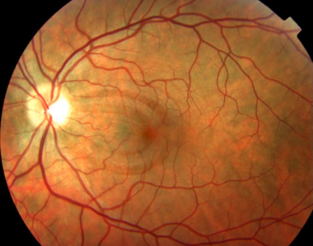
Woman referred for blurry vision, flashes of light and floaters

Nerve Fiber Analysis - Glaucoma Associates of Texas

Sample of retinal nerve fiber layer (RNFL) report provided by